Reader
Reader displays protein structures and trajectories alongside calculated properties. The examples below show atom selection, geometry measurements, shielding tensors and plots of quantities across frames.
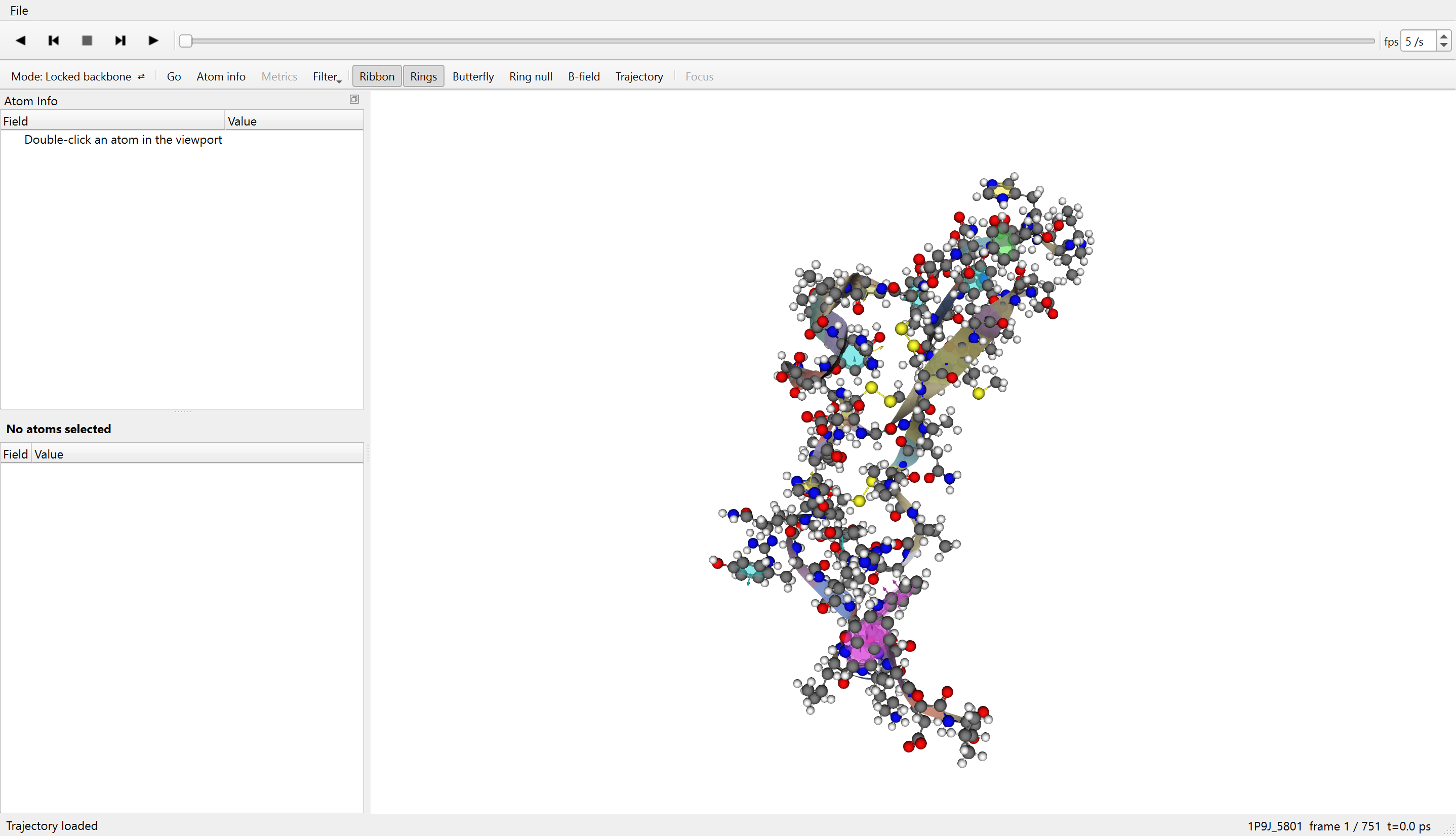
1P9J in Reader. Playback controls are above the structure. Atom Info and the measurement and tensor readouts share the panel on the left.
Installation
Windows
Reader is available as a Windows 64-bit installer. A 100-frame example is included; the other trajectories can be selected in the application and downloaded separately.
The installer is not code-signed. Windows may display an unrecognised-app warning. The download is approximately 1.08 GiB; the browser handles download progress and any interruption.
macOS
Open the DMG and drag H5 Reader to Applications.
Trajectory catalogue
Open an .lgs file from disk, or choose File → Open from website. A collection file opens a searchable list; an individual run opens the protein. The catalogue includes 173 production trajectories and three more densely sampled trajectories: 1P9J, Trp-cage and chignolin.
The catalogue supports searches by protein name, PDB or BMRB identifier, organism and other listed keywords. Reader copies packages from a drive or downloads them from the website into a local buffer, then unpacks them before opening. Source files are unchanged.
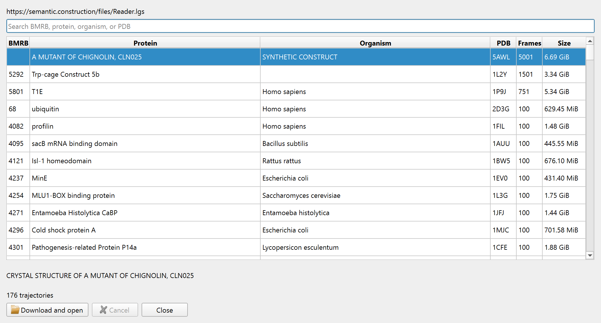
The same collection dialogue serves disk and web sources. Package sizes are shown before transfer. A web download can be cancelled and resumed with Retry.
Atom selection
Double-click an atom in the structure, or use Go to find it by name. Atom Info identifies the atom, residue and chain, and lists the available values for the current frame. Names in the supported naming conventions appear together.
Filter limits the view to selected residues. The example shows Tyr46's sidechain and carbonyl group with the rest of the protein hidden. Filtering changes the display, not the stored structure.
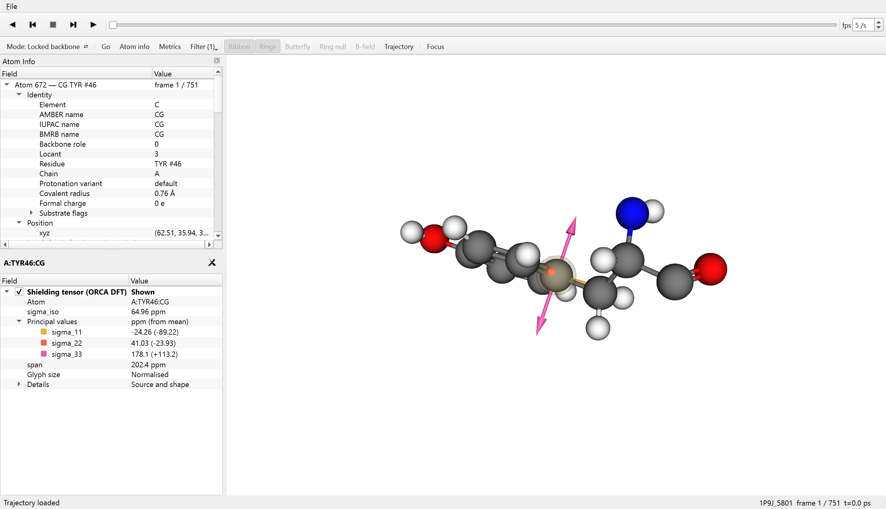
Tyr46, CG. Atom Info lists the selected atom's identity and values for the current frame. An ORCA shielding tensor is available for this atom.
Geometry measurements
Hold Shift while double-clicking more atoms. Two atoms give a distance, three an angle, and four a dihedral. The readout lists the atoms in selection order and identifies the angle's vertex or the dihedral's axis. It updates when the frame changes.
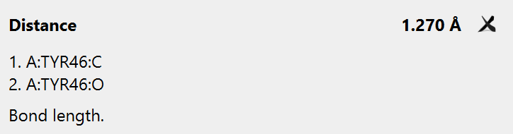
Two atoms: C–O bond length.
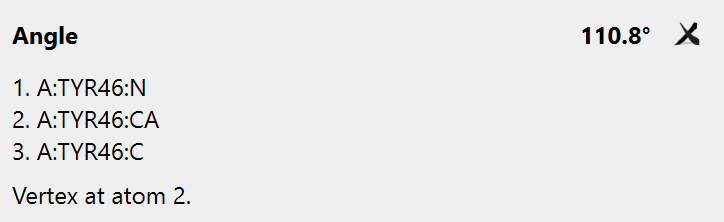
Three atoms: N–CA–C angle.
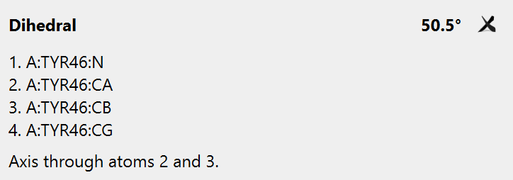
Four atoms: the sidechain χ1 dihedral.
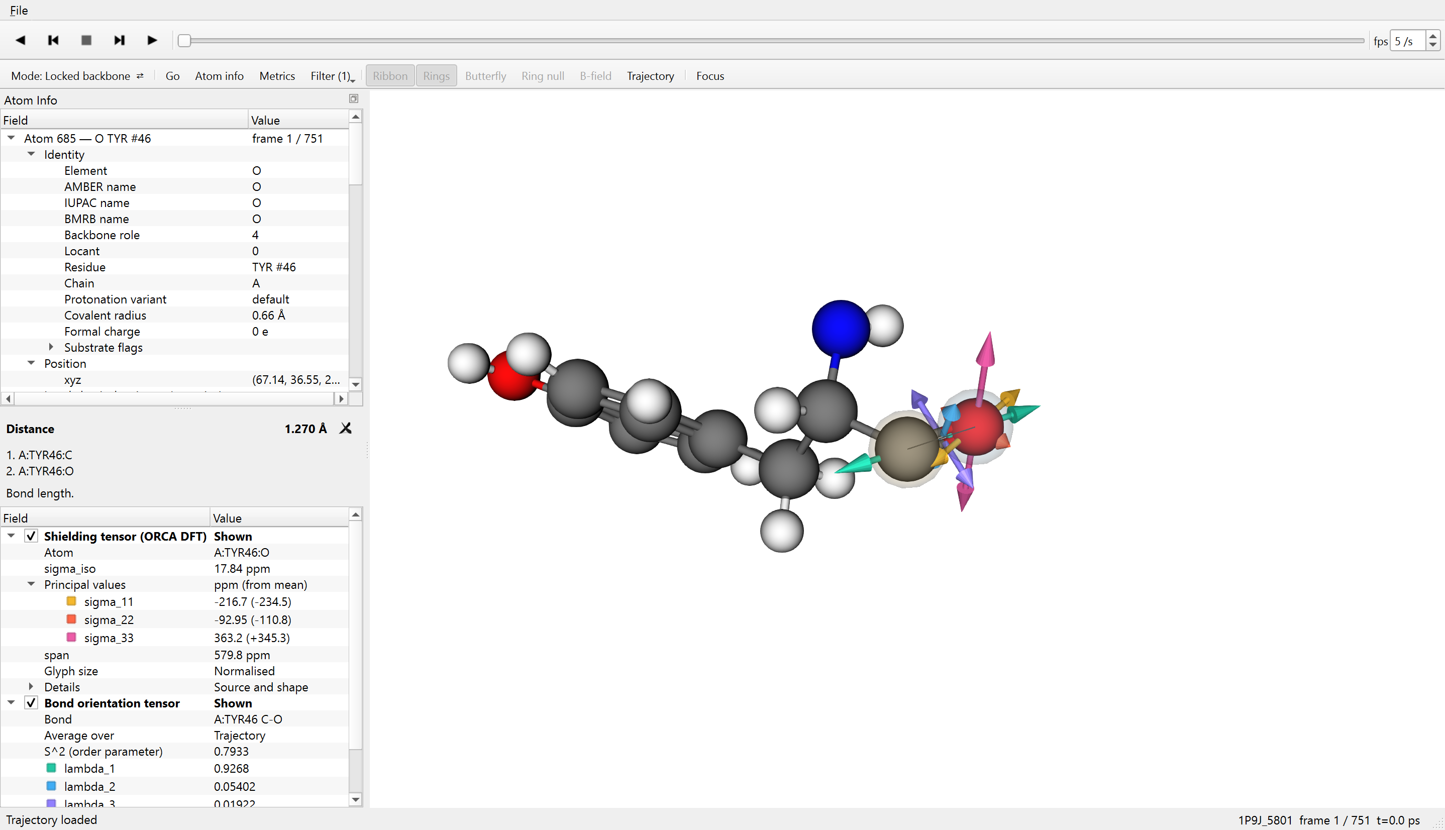
The measured C–O distance is 1.270 Å in this frame.
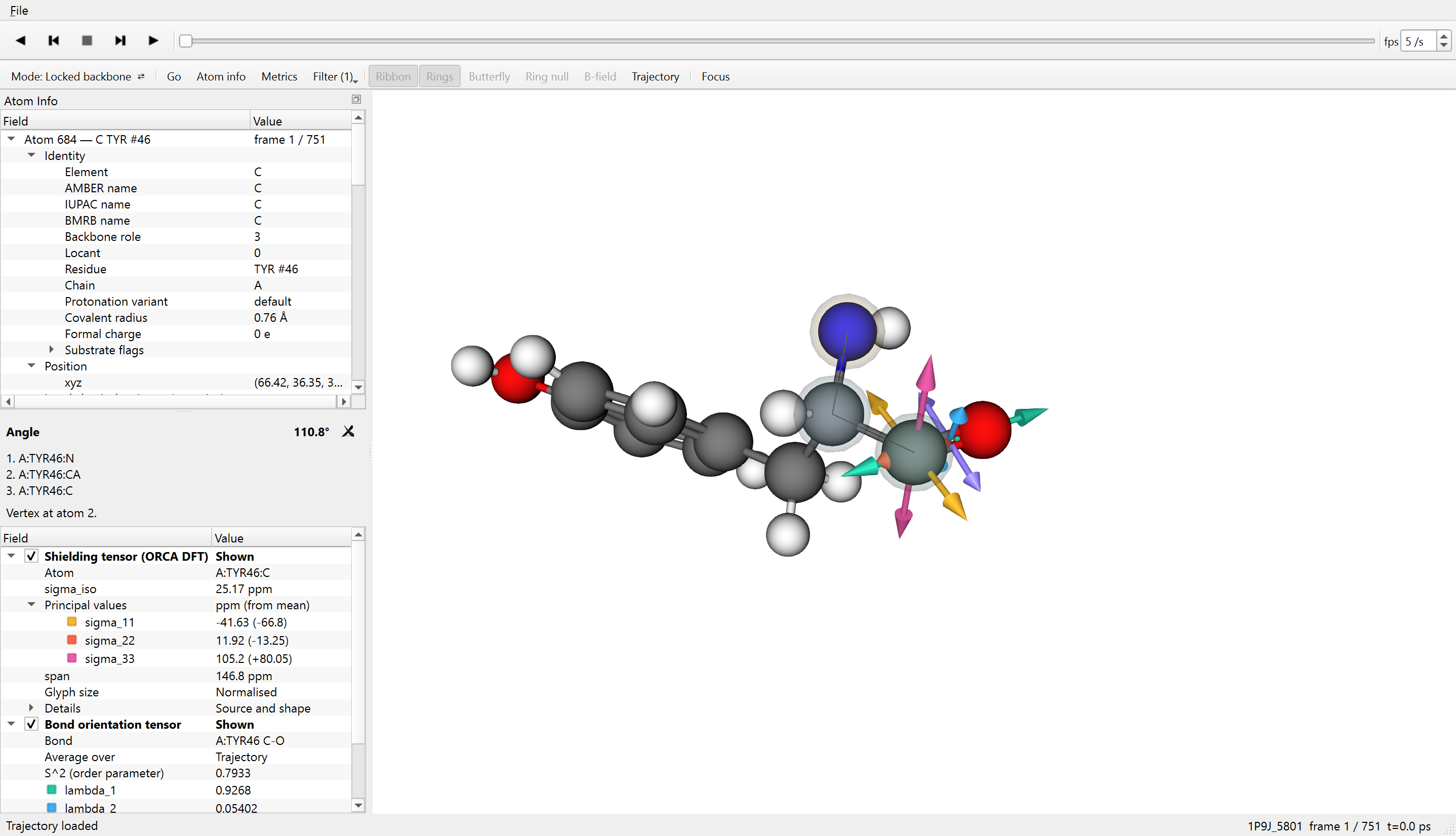
The N–CA–C angle is 110.8°. CA, the second selected atom, is the vertex.
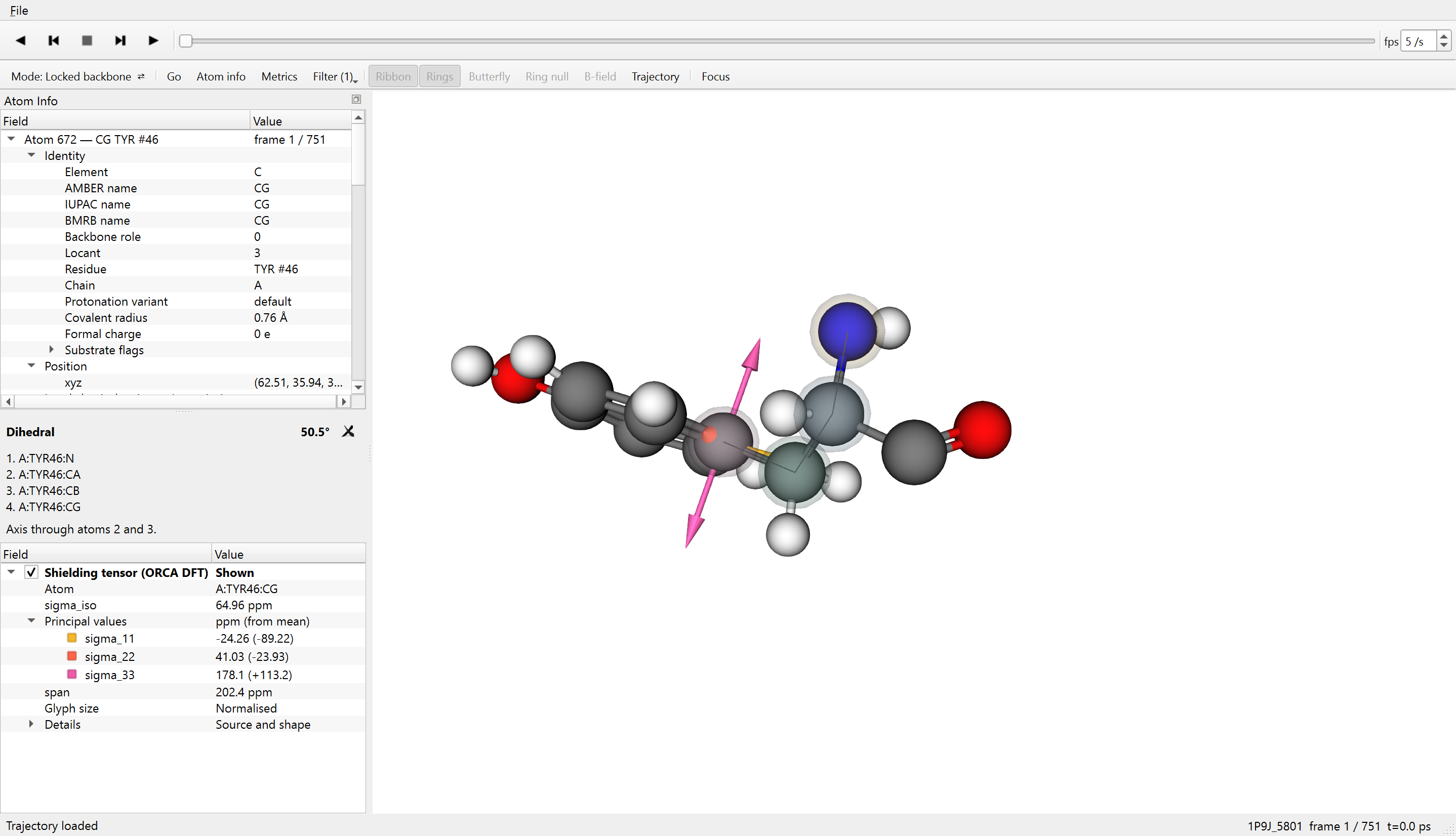
The N–CA–CB–CG dihedral is 50.5°. The line follows the selected atoms; the rotation axis runs through CA and CB. The pink arrows belong to the selected atom's shielding tensor.
Tensor display
A shielding tensor describes how an atom's magnetic shielding depends on direction. Reader draws its principal axes at the atom and lists the principal values in the panel. Colours match each value to its axis in the structure.
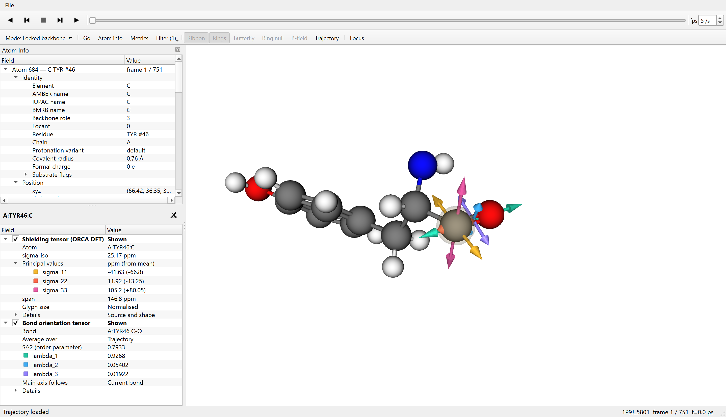
Shielding and bond orientation at Tyr46's carbonyl group. Warm colours identify shielding; cool colours identify bond orientation. Each tensor has a checkbox to show or hide it. With both unchecked, the atom remains selected without either tensor displayed.
Shielding at the selected frame
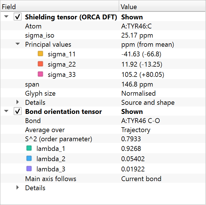
The ORCA result here has an isotropic mean, σiso, of 25.17 ppm. Its principal values are −41.63, 11.92 and 105.2 ppm. Parentheses in the panel give each value's difference from the mean.
Reader normalises each glyph separately, so its overall size does not provide a common scale for comparing atoms. The panel provides the numerical shielding values in ppm, not experimental chemical shifts.
Bond orientation across the trajectory
The bond-orientation tensor summarises how the C–O direction varies across the aligned frames. Its three values are mean squared projections onto its principal axes; they sum to one. The panel also reports the order parameter, S2.
For display, Reader places this glyph with its main axis along the current bond. Its shape summarises the trajectory; its displayed orientation is not the orientation of the average tensor in the aligned protein.
Tensor values: enlarged panel
Predicted shielding
On a compatible run without an ORCA tensor, selecting an atom requests a prediction from Reader's bundled experimental model. The same tensor panel displays the result, now labelled Predicted.
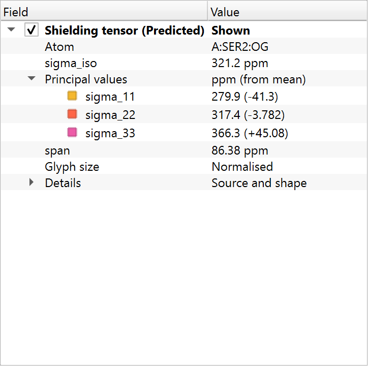
This example comes from the 100-frame trajectory for BMRB 4095, sacB mRNA binding domain. Reader predicts the shielding of Ser2's sidechain oxygen from the extracted molecular features. There is no attached ORCA calculation in this package.
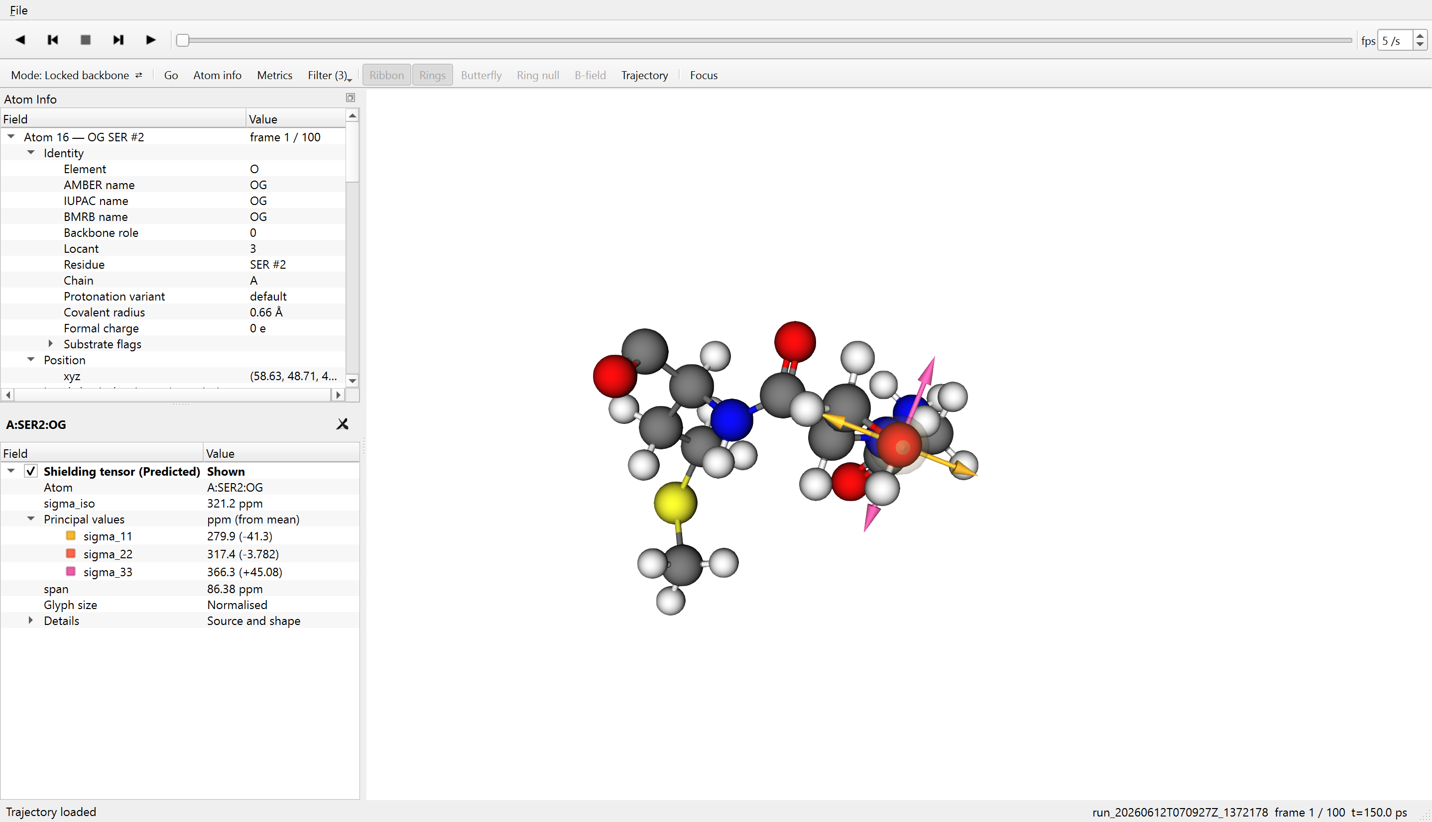
Ser2, OG, first displayed frame (150 ps). The predicted isotropic shielding is 321.2 ppm. Principal values are 279.9, 317.4 and 366.3 ppm.
Predicted values: enlarged panel
Field definitions
Right-click a field in Atom Info or a metric in the Metrics dialogue to read its meaning, calculation and origin. The glossary distinguishes quantities calculated by nmr_extract, results from tools such as ORCA and DSSP, and values calculated by Reader.
The Metrics dialogue lists quantities for the selected atom, its residue, bonds and nearby aromatic rings. The list is searchable, and the available display choices depend on the quantity.
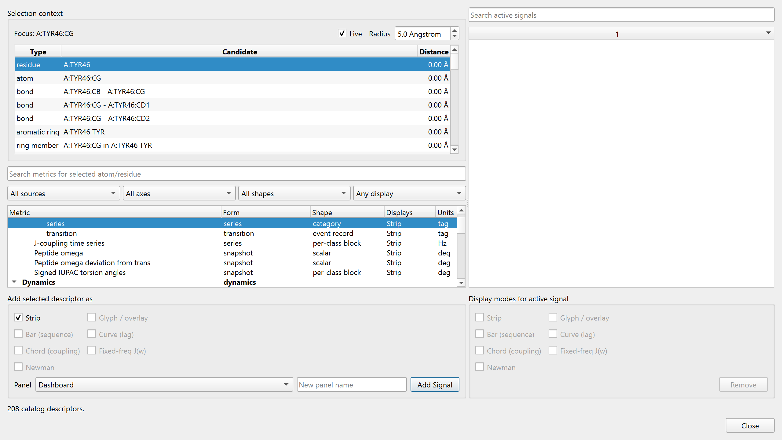
Metrics around Tyr46. Choose the object of interest at the top left, then a quantity and its display below. Selected signals are listed on the right.
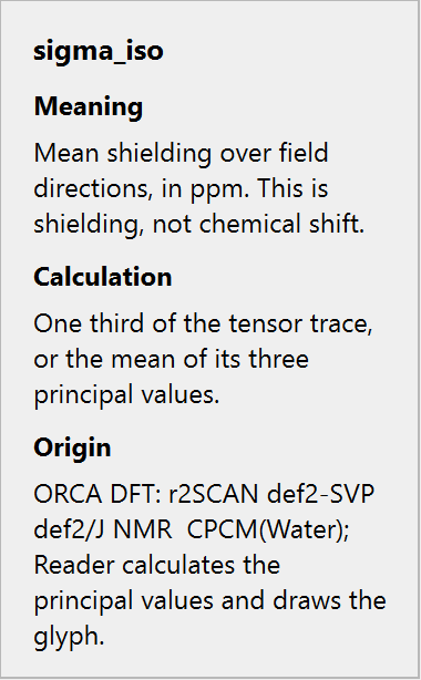
Right-click help for isotropic shielding. The popup explains the quantity, how it is obtained, and which calculation supplies it. Click outside it to return to the structure.
Plots across frames
Add a quantity as a Strip in Metrics to plot it across frames. Playback and the frame slider update the molecular view, allowing changes in the trace to be compared with the corresponding structures.
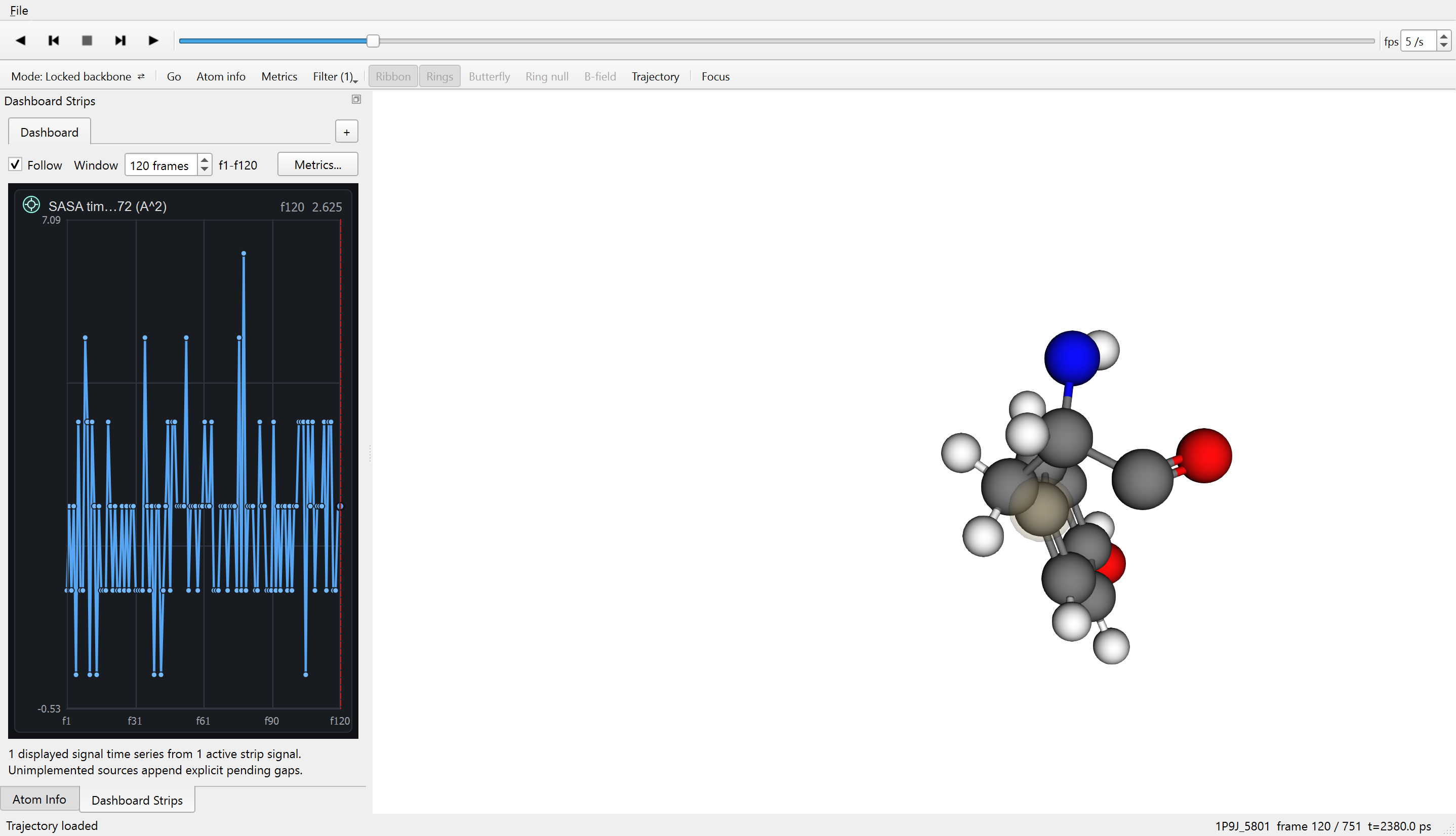
SASA of Tyr46 CG over 120 displayed frames. The plot is in Å2; the structure is at frame 120, 2,380 ps. The trace shows the stored surface-area values as the sidechain moves.
Depending on the run, the available quantities include dihedrals, secondary structure, solvent exposure, hydrogen-bond geometry, electronic descriptors and shielding. The catalogue and source labels identify what is present in the file you opened.